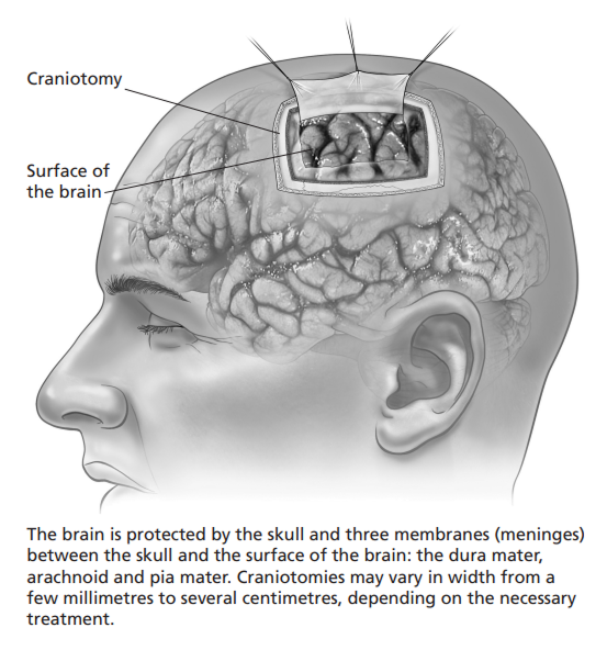
What is a Craniotomy?
A craniotomy is a procedure to open temporarily a part of the skull to expose the brain for surgery. Advances over the past 20 years have made craniotomy safer, simpler and more successful. Neurosurgeons can now operate in areas of the brain that once were thought impossible to reach. Craniotomy is performed as a part of the surgical treatment of many different conditions, including: • a growth within the brain or from membranes that surround it. • a blood clot pressing on the brain, often from an acute head injury. • weakness in an artery, a cerebral aneurysm. An aneurysm forms when the weak spot bulges. In some patients, the aneurysm may enlarge, increasing the risk that it may rupture and cause major bleeding in the brain. Many aneurysms can be treated with procedures performed through the leg. In some cases, a neurosurgeon may place a titanium clip near the aneurysm to seal it from the normal artery
An untreated aneurysm may burst and cause life-threatening bleeding into the brain.
Diagnosis
Diagnostic Imaging
Advanced imaging techniques and improved computer technology have led to more accurate diagnosis, exact locating of the problem, and precision surgery. The diagnostic imaging tests most commonly used prior to neurosurgery are: magnetic resonance imaging (MRI), computer tomography (CT), angiography and X-ray examination. One or more of these tests may be necessary.
A decision whether to have surgery
As you make the decision whether to have surgery, make sure that you understand the risks, benefits and limitations of surgery. If you do not have surgery, your symptoms and condition may continue to worsen. Only you can decide if surgery is right for you. If you have any questions, ask your surgeon.